When you first hear about open globe injuries, you might wonder what makes them so critical and how they differ from other eye injuries. With their potential to cause severe pain and even permanent vision loss, these injuries don’t just affect your sight—they can alter your life. But what exactly leads to these traumatic incidents, and why are they more common in certain demographics? Understanding the symptoms, risk factors, and treatment options becomes essential. As you explore this topic further, you’ll uncover the steps needed to protect and possibly restore vision after such a devastating event.
Key Takeaways
- Open globe injuries are full-thickness wounds of the eye wall, often caused by sharp objects, falls, or violent encounters.
- Symptoms include severe eye pain, vision loss, fluid leakage, and visible ocular injuries.
- Non-contrast CT scans are preferred for damage assessment and identifying foreign bodies.
- Immediate surgical intervention within 24-48 hours is crucial for optimal visual recovery.
- Prevention includes securing sharp objects, using protective eyewear, and education on safe tool handling.
Understanding Open Globe Injuries
Open globe injuries (OGIs) seriously threaten ocular health and demand immediate attention and intervention. These full-thickness wounds of the eye wall include globe rupture, laceration, penetration, and perforation. With an incidence rate of about 3.5 injuries per 100,000 people annually, understanding OGIs is vital. They often result from eye trauma due to falls, sharp objects, vehicle accidents, or violent encounters.
The initial evaluation is essential When dealing with an open globe injury. It helps determine the extent of the damage and the presence of any intraocular foreign body. This step sets the stage for surgical intervention, which should occur within 24-48 hours. Such timely intervention is important to optimize visual outcomes and minimize complications.
The prognosis largely depends on the injury’s initial mechanism and how quickly medical care is provided. For instance, a penetrating injury with an intraocular foreign body might pose more severe risks than other types.
The Ocular Trauma Score can be a valuable tool for predicting visual outcomes. It can also help the medical team, and you understand the potential recovery trajectory. Act fast; your vision might depend on it.
Symptoms and Risk Factors
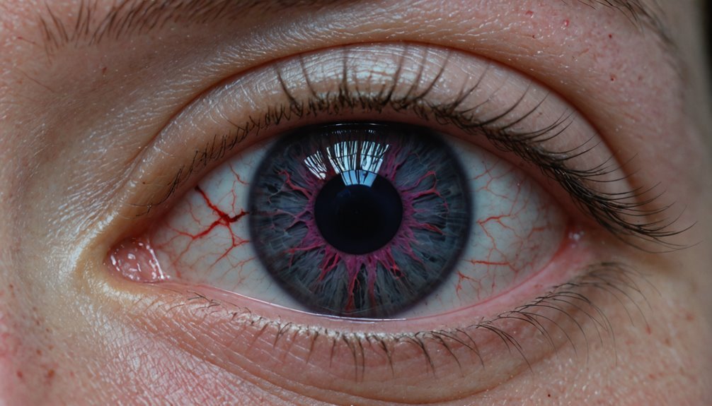
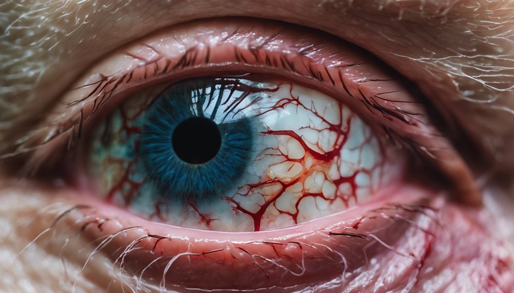
Experiencing an open globe injury can be alarming, with symptoms that demand immediate attention. You might notice severe pain, vision loss, and other distressing signs. Fluid could leak from the eye, and you might see visible injuries to the eyelid or even extrusion of ocular tissue.
These symptoms aren’t just uncomfortable—they’re urgent signals to seek medical help immediately.
Understanding the risk factors for open globe injuries can help you stay vigilant. Common causes include sharp object injuries, particularly in children, and falls, especially in individuals over 75.
Other significant risk factors include:
- Previous eye surgeries might’ve weakened the eye’s protective tissues.
- Activities involving sharp tools or high-velocity projectiles.
- The age group most affected tends to be males, particularly those between 10 to 30 years.
- Men and boys account for about 80% of these cases.
Open globe injuries also result from vehicle accidents and violent encounters. The incidence rate is about 3 per 100,000 people in the U.S., but it’s higher in developing countries due to less stringent safety measures.
Staying informed and cautious can be vital in preventing these potentially life-changing injuries.
Diagnostic Techniques
When approaching open globe injuries, start with a thorough eye examination that includes visual acuity assessment and pupillary response testing to gauge initial functionality.
Non-contrast CT scans are used to evaluate the extent of damage effectively and identify any intraocular foreign bodies.
Don’t overlook the importance of slit-lamp examinations and B-scan ultrasonography to spot less obvious injuries while considering differential diagnoses like traumatic hyphema and orbital fractures.
Eye Examination Essentials
A thorough eye examination is essential for accurately diagnosing and effectively managing open globe injuries.
You’ll need to start with an eye examination, measuring visual acuity to assess how well the patient sees. Carefully check the pupillary response, as changes can indicate severe trauma. Remember, a gentle approach is important to avoid further damage.
Use a slit lamp to evaluate the anterior segment of the eye. This tool helps you identify any lacerations or foreign bodies that might be present. It’s a significant step in understanding the full scope of the injury.
After the slit lamp examination, imaging techniques come into play. A non-contrast CT scan is recommended to confirm the diagnosis and determine the extent of the damage.
Here are essential steps to follow during the examination:
- Visual acuity measurement: Determine the patient’s baseline vision.
- Pupillary response assessment: Check for any irregularities indicating neurological issues.
- Slit lamp examination: Inspect the eye thoroughly for foreign bodies and lacerations.
- CT scan: Utilize to clarify the injury’s severity and guide further treatment plans.
Document your findings thoroughly; this information is important for coordinating with ophthalmology specialists for ongoing care.
Imaging Methods Overview
Evaluating open-globe injuries demands precision, and imaging methods play a vital role. When faced with such a challenge, non-contrast CT scans are preferred. They effectively detect associated injuries with an accuracy of 81% and a sensitivity of 76%, which is better for planning any surgical intervention. However, if you’re dealing with a case with limited direct examination, B-scan ultrasonography becomes invaluable. It excels at identifying intraocular foreign bodies (IOFBs) and evaluating posterior segment injuries.
Here’s a quick comparison of imaging methods:
| Imaging Method | Key Features |
|---|---|
| Noncontrast CT | Preferred, 81% accuracy, 76% sensitivity |
| B-scan Ultrasonography | Identifies IOFBs, evaluates posterior injuries |
| MRI | Contraindicated for open-globe injuries |
Remember that MRI is contraindicated due to the risk of ferromagnetic materials and its insensitivity to foreign objects. As you evaluate these injuries, remember that imaging isn’t just about diagnosis. It’s also about documenting findings to guide treatment decisions and guarantee clear communication among the care team. Proper documentation aids in strategizing surgical interventions while avoiding further damage to surrounding structures.
Identifying Injury Indicators
Visual acuity and pupillary response must be evaluated to diagnose open globe injuries accurately and establish a baseline.
During this initial evaluation, look for common signs such as thick subconjunctival hemorrhage and a peaked pupil, which might indicate a globe rupture or laceration. Identifying these signs early can guide further diagnostic steps and treatment.
Non-contrast CT scans are the best diagnostic imaging tool when you suspect a serious injury. They are essential for confirming a globe rupture and determining the injury’s extent, with an accuracy rate of 81% and a sensitivity of 76%.
However, don’t stop there. A thorough slit-lamp examination is essential to detect intraocular foreign bodies and other internal damage. If direct examination is challenging, consider using ultrasound for a better look.
Here’s what you need to focus on:
- Visual acuity and pupillary response: Establish a baseline.
- Subconjunctival hemorrhage: Look for signs of rupture.
- Diagnostic imaging: Utilize CT scans for accurate evaluation.
- Surgical exploration: Act swiftly to prevent complications.
Immediate surgical exploration is often necessary for suspected globe ruptures, as delays can lead to complications like choroidal hemorrhage.
Treatment and Management
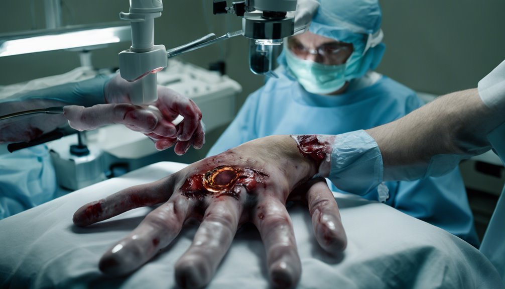
Open globe injuries demand immediate attention and surgical intervention within 24-48 hours to guarantee the best visual outcomes and minimize complications. As soon as you’re faced with an eye injury of this nature, the initial management steps are fundamental.
Begin by stabilizing the patient’s overall condition and shielding the eye to prevent further damage. Administer prophylactic antibiotics right away to ward off posttraumatic endophthalmitis, a severe infection that can lead to devastating consequences.
Diagnostic imaging, especially non-contrast CT scans, helps assess the full extent of the injuries, guiding your surgical planning effectively.
After surgery, the focus shifts to vigilant postoperative care. Topical antibiotics and corticosteroids should be applied to prevent infections and inflammation.
Regular follow-up exams are essential to monitor potential complications like retinal detachment and endophthalmitis. The Ocular Trauma Score comes into play by providing a predictive visual prognosis based on the initial characteristics of the injury.
Remember, low scores generally indicate poorer visual outcomes, emphasizing the significance of swift and efficient management.
Maintaining clear communication with the patient about the steps being taken and expected outcomes is important for effective treatment and management throughout this process.
Prognosis and Recovery
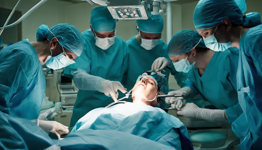
When facing an open globe injury, acting swiftly is vital, as early surgical intervention within 24-48 hours can greatly improve your chances for visual recovery.
Your long-term vision outcomes will depend on factors like the severity of the injury and the initial treatment. Many people achieve functional vision through prompt care.
Postoperative care is essential, so you’ll need regular follow-ups to address potential complications and adhere to guidelines like avoiding strenuous activities for six weeks.
Early Intervention Importance
Timely intervention in open globe injuries is essential for optimizing visual outcomes. Acting quickly, ideally within 24-48 hours, markedly reduces the risk of irreversible damage and complications.
Early intervention is vital in managing open globe injuries, especially when dealing with a globe rupture, which can severely impact prognosis. Immediate surgical repair and antibiotic prophylaxis help prevent posttraumatic endophthalmitis, which affects 3-10% of cases.
Without timely surgical intervention, complications such as retinal detachment and proliferative vitreous hemorrhage become more likely, threatening your chance of preserving vision.
Consider these important points:
- Initial Visual Acuity Matters: The Ocular Trauma Score indicates that your initial visual acuity greatly influences long-term recovery.
- Severity of Injury: A ruptured globe markedly worsens prognosis, emphasizing the need for swift treatment.
- Complication Risks: Early repair minimizes the risks of retinal detachment and other severe complications.
- Antibiotic Prophylaxis: Prompt use of antibiotics reduces the likelihood of posttraumatic infections.
Long-term Vision Outcomes
Long-term vision outcomes after an open globe injury hinge on several vital factors, with initial visual acuity being a key determinant. Your recovery prospects are generally more favorable if your vision was relatively good before the injury.
The Ocular Trauma Score (OTS) is a valuable tool for predicting visual prognosis. A higher OTS suggests a better chance for vision recovery, helping set realistic expectations for you.
Complications pose significant challenges to long-term vision recovery. Retinal detachment, endophthalmitis, and intraocular foreign bodies can severely impact outcomes.
Endophthalmitis, a serious infection, occurs in 3% to 10% of penetrating injuries, underscoring the need for prompt care. Surgical intervention within 24-48 hours of the injury is essential. Quick action minimizes the risk of complications and optimizes the chances of restoring vision.
Your journey to recovery doesn’t end with surgery. Follow-up care is essential for addressing ongoing issues like secondary cataracts or glaucoma.
Regular monitoring and potential additional treatments can help manage these conditions effectively. Staying proactive and adhering to follow-up care increases your chances of achieving the best possible long-term vision outcomes after an open globe injury.
Postoperative Care Guidelines
Following surgery for an open globe injury, adhering to postoperative care guidelines is vital for ideal recovery and prognosis. You’ll need to wear a protective eye shield for several weeks to protect your eye and guarantee proper healing. This prevents pressure and further injury to the eye.
Avoid eye rubbing, heavy lifting, exercise, and swimming for at least six weeks. These activities can jeopardize your recovery and visual acuity.
Regular follow-up examinations are important for monitoring your progress. During these check-ups, your doctor will assess your visual acuity and look for potential complications, such as secondary cataracts or retinal detachment. These complications can greatly impact your recovery and prognosis.
The Ocular Trauma Score (OTS) helps predict your visual outcomes, relying on your initial visual acuity and any complications you may encounter.
To minimize infection risks, prophylactic antibiotics are usually administered for two days post-surgery. This is essential as posttraumatic endophthalmitis occurs in 3 to 10% of penetrating trauma cases.
By closely following your postoperative care plan, you can maximize your chances of a successful recovery.
- Use a protective eye shield
- Avoid certain activities for six weeks
- Attend regular follow-up exams
- Take prescribed prophylactic antibiotics
Prevention Strategies
In preventing open globe injuries, securing sharp objects and keeping them out of reach, especially from children, is fundamental. Young boys aged 10-30 are particularly at risk.
Prevention is key, and wearing protective eyewear during hazardous activities or in risky environments can greatly reduce the chances of eye injuries. Many open-globe injuries occur during sports or work-related tasks, so don’t underestimate the power of protective gear.
Education plays an essential role, too. Teaching individuals how to handle tools and equipment safely can mitigate risks, particularly in occupational settings. Awareness of one’s surroundings and understanding potential hazards can lead to quicker responses if an eye injury occurs.
Regular eye exams are important for everyone, especially if you have pre-existing conditions that could worsen the impact of an injury.
Occupational safety measures should be prioritized in workplaces to guarantee that everyone understands the importance of eye protection and safe practices.
Frequently Asked Questions
What Is an Open Globe Injury?
You’ve encountered an eye injury involving trauma and visual impairment. Understand the eye anatomy to assess damage. Emergency care is vital, and surgical techniques help the patient recover. Be aware of risk factors and guarantee eye protection to prevent future incidents.
How Do You Repair an Open Globe?
You repair an open globe using surgical techniques to close the wound, address intraocular pressure, and promote visual rehabilitation. To guarantee an ideal recovery, you administer antibiotic therapy, provide follow-up care, and educate patients on risk factors.
Is a Ruptured Globe an Emergency?
Yes, a ruptured globe is an emergency. Start by immediately assessing visual acuity and risk factors. Then, prioritize patient stabilization, diagnostic imaging, and surgical intervention. Finally, it guarantees proper follow-up care to address complications and optimize recovery.
What Is the Prognosis for a Ruptured Globe?
You should understand that the prognosis depends on initial visual acuity and surgical interventions. Treatment options and patient education are essential. Long-term effects and recovery timelines vary, requiring thorough risk assessment for better visual outcomes.
Conclusion
In dealing with open globe injuries, you must act quickly. Recognizing symptoms and understanding risk factors can help you seek timely medical attention, which is critical for preserving vision. Diagnostic techniques are there to pinpoint the injury, and treatment should ideally occur within 24-48 hours to reduce complications. Recovery varies, but prompt intervention improves outcomes. Remember, prevention strategies can greatly lower your risk of these severe injuries, so always prioritize eye safety.
